Clinical Insights
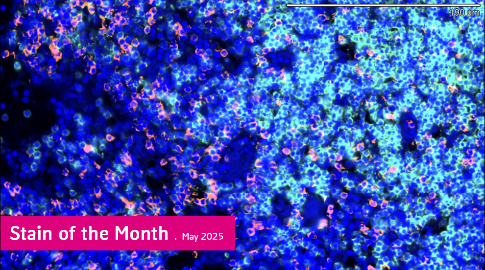
CD4/CD8 Duplex Immunofluorescence:
High-Resolution Immune Profiling from a Single Slide
by Laura Kurth, PhD, VP of Laboratory Operations
This month we’re highlighting our CD4/CD8 Duplex Immunofluorescence assay as the Stain of the Month for its utility in visualizing and quantifying T-cell subsets in tissue.
The assay enables simultaneous detection of CD4⁺ T-helper cells and CD8⁺ cytotoxic T cells using spectrally distinct fluorophores. It is fully automated, tissue-sparing, and optimized for use on formalin-fixed paraffin-embedded (FFPE) samples, making it ideal for both prospective studies and retrospective tissue analysis. This approach provides reproducible, high-resolution insight into immune cell behavior and spatial distribution in clinical and research contexts.
Why CD4 and CD8?
CD4⁺ T cells coordinate immune responses via MHC class II and serve as key regulators of adaptive immunity. CD8⁺ T cells interact with MHC class I to eliminate infected or malignant cells. Analyzing both populations in situ reveals critical information about immune engagement, suppression, and tissue context.
Why It Matters in Preclinical Research
Quantifying CD4+ and CD8+ T-cell subsets in tissue is vital to understanding how investigational drugs modulate the immune system. This is particularly critical in the preclinical phase, where visualizing immune cell infiltration, activation, and localization provides answers to:
1. Is the drug engaging its target in tissue?
2. Is there a shift in the immune microenvironment?
3. How does immune cell distribution relate to tumor regression or tissue pathology?
Key Advantages
- Simultaneous dual-marker detection enables co-localization and comparative quantification of CD4⁺ and CD8⁺ cells on the same slide
- Fully automated workflow reduces variability, increases throughput, and ensures consistency across batches
- High-resolution, fluorescence-based imaging enhances visualization of immune subsets, even in dense or complex tissue regions
- Optimized for FFPE samples, including limited biopsies, making it ideal for retrospective studies and tissue-conserving protocols
- Ready-to-integrate into clinical trial pipelines, with validated protocols and digital data outputs available on request
Applications
- Immuno-oncology
Assess tumor-infiltrating lymphocytes (TILs), immune exclusion, and CD4/CD8 ratios in the tumor microenvironment. - Transplant pathology
Monitor T cell-mediated rejection and immune activation in graft tissue. - Autoimmune disease research
Evaluate T-cell distributions and imbalances in inflamed organs such as kidney, skin, and salivary glands. - Clinical trials
Stratify patients, assess immunomodulatory effects, and support immune biomarker development.
Digital Pathology Integration
To extend the value of our CD4/CD8 duplex immunofluorescence assay, MLM offers integrated AI-powered digital pathology tools. These tools enable precise, automated classification of CD4⁺ and CD8⁺ T cells directly within complex tissue environments. With advanced image analysis, we can quantify immune cell densities, calculate CD4:CD8 ratios within defined regions of interest, and map the spatial distribution of T-cell populations across tumor, stromal, or inflamed areas.
In addition, proximity analysis can be used to examine the relationships between immune cells and their targets—helping researchers explore functional immune dynamics within the tissue microenvironment. All results are available as structured, exportable data sets, making this solution ideal for biomarker development, publications, and regulatory submissions. This digital integration enhances the reproducibility, depth, and scalability of immune profiling—especially in high-throughput or multicenter study settings.
Supporting Translational Research
Currently used in oncology and immunology-focused studies, this assay is ready to support your immune profiling objectives—from early-phase research to clinical trial deployment.
To learn more or request technical specifications and sample images, get in touch with our team today: