Clinical Insights
Reticulin Staining:
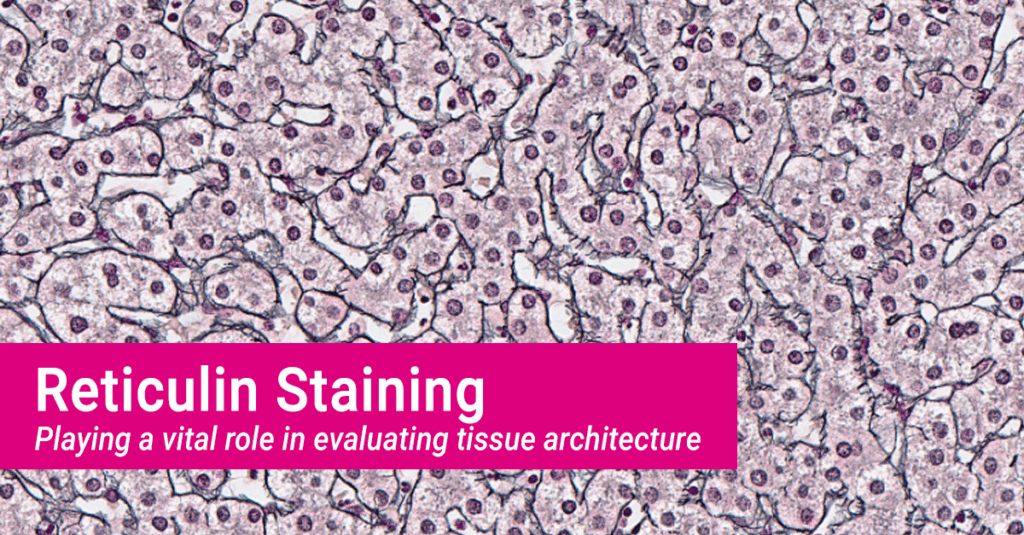
Playing a vital role in evaluating tissue architecture
by Laura Kurth, PhD, VP of Laboratory Operations
MLM Medical Labs highlights Reticulin Staining for its ability to inform on assessment of tissue architecture.
Reticulin staining is a histochemical method used to selectively visualize type III collagen fibers, which form a fine reticular framework supporting parenchymal cells in organs such as the bone marrow, liver, spleen, and lymph nodes. These fibers are not detectable with routine hematoxylin and eosin (H&E) staining but become clearly visible using silver impregnation techniques—most notably the Gordon & Sweets reticular fiber method. This stain plays a vital role in evaluating tissue architecture, detecting early stromal changes, and identifying pathologic patterns of fibrosis or infiltration.
Core Diagnostic Applications
- Structural Evaluation: The reticulin network outlines the normal lobular or trabecular organization in complex tissues. Disruption of this framework indicates architectural distortion caused by neoplasia, chronic damage, or regenerative remodeling.
- Fibrosis Identification: Reticulin staining is particularly sensitive to early fibrotic changes that precede collagen deposition detectable by trichrome stains. This is valuable in evaluating chronic liver disease, bone marrow fibrosis, and reactive stromal responses.
- Lesion Differentiation: A well-preserved reticulin framework is typically seen in benign lesions (e.g., hepatocellular adenoma), whereas malignant neoplasms (e.g., hepatocellular carcinoma) often show disruption, thickening, or loss of the reticulin mesh.
- Detection of Tumor Infiltration: In settings such as bone marrow biopsies or lymph node evaluations, reticulin staining helps reveal infiltrative disease, including hematologic malignancies and metastatic tumors, by highlighting effacement or remodeling of the stromal network.
Clinical Relevance
The Gordon & Sweets technique provides a reliable and reproducible method for assessing the reticulin framework. Despite the rise of immunohistochemistry and molecular diagnostics, reticulin staining remains a cost-effective, rapid, and high-yield adjunct that offers essential structural information. It continues to be integral in:
- Detecting early or subtle fibrosis
- Assessing tissue invasion or remodeling
- Supporting histologic interpretation when other stains are inconclusive
Despite advances in molecular diagnostics, reticulin staining continues to provide irreplaceable architectural context across numerous tissue types. As part of our comprehensive histology services, MLM Medical Labs ensures each stain delivers clarity, consistency, and value in every case. We are committed to delivering scientifically rigorous data that enhances your diagnostic confidence.